Imaging through a kaleidoscope

The first kaleidoscope was developed in the early 1800s by Sir David Brewster, who was seduced by the beauty of the generated patterns, which are symmetrical and, at the same time, very complex. In our study, we demonstrate that the kaleidoscopic effect, beyond its artistic function, can be exploited by scientists working with optical fibers.
The development of optical endoscopes is motivated by a number of biomedical applications such as brain imaging. Multimode fibers are excellent candidates to minimize the invasiveness of these procedures. However, the propagation of light in multimode fibers is very complex. Indeed, in this type of fiber, light propagates in an unpredictable way, producing patterns that are difficult to interpret at the fiber output. Techniques currently exist to reconstruct images from such patterns, but these techniques are very sensitive to all deformations applied to the fiber, which strongly limits the applicability of these techniques in biology.
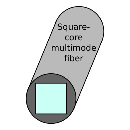
In our study, we demonstrate that it is possible to produce images with multimode fibers even if they are deformed. To this end, we were inspired by Sir David Brewster's kaleidoscope, and we traded our usual fibers, which have a circular core, for square-core fibers. Indeed, the symmetry properties of these fibers generate a remarkable kaleidoscopic effect, which transports information in a way robust to deformations. We tested this method by reconstructing images of fluorescent sources through the fiber, even when this fiber was randomly deformed, thus reproducing the dynamic aspect of the perturbations that typically occur when studying living organisms. Despite these perturbations, we successfully managed to reconstruct images of the sources through the fiber. This new technique is thus promising for the development of miniature endoscopes for biomedical imaging.
This study has been published in PNAS (Bouchet et al., 2023).
